Birth of the first Epidermis Atlas, an advance in research into herbivore diets
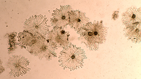
A UAB team has developed a Digital Plant Epidermis Atlas, the first consultation tool of its type to exist until now, which will allow to advance in the research of the diets of herbivores. This open database of images shows the microanatomic characteristics of 400 plant species obtained through optical microscope.
22/10/2020
As part of the conference on wild ungulates of the Iberian Peninsula entitled XI RUSI (Reunión de Ungulados Silvestres Ibéricos), organised in Madrid as an online conference on 23 and 24 October with the participation of experts and researchers in these animal species, a UAB team presented the digital epidermis atlas, developed in the past year by a team formed by biologists (Jordi Bartolomé, Omar López and Emmanuel Serrano) and members of the Library Services (Vicenç Allué and Cristina Andreu).
No such tool had existed until now, and it was designed as a way to contribute relevant improvements in research through the microhistological analysis of the diets of herbivore species.
The microhistological analysis of plant epidermis in the faeces and stomach contents of wild hoofed animals (ungulates) to determine their diet is something that has been done for decades. Although new techniques have appeared, such as molecular or spectrophotometric analysis, the microhistological method continues to be the one to provide the greatest amount of information in these types of studies. It is nevertheless an arduous method that requires the creation of plant epidermis collections.
The possibility of having an open-access photographic database available to all will speed up these types of analyses. The atlas is located in an institutional archive, the Digital Deposit of Documents of the UAB (DDD), which guarantees long-term availability and preservation. The link to the altas is https://ddd.uab.cat/collection/atlepi?ln=en.
The database of images shows the microanatomic characteristics of plant epidermis of some 400 species, obtained using an optical microscope. It offers details on the cell shapes, stomas, and trichomes at 100 and 400 magnification. The atlas covers many of the most common species in the Mediterranean region, and the goal is to expand the atlas with species from other regions.
With this in mind, the team behind the altas aims to present it as a consultation tool for researchers using microhistological analyses in their studies to identify plant debris in the faeces and stomach contents of herbivores. The team invites researchers from all over the world to add more images to the atlas in order to make the database grow.
In a second phase, starting in 2021, the researchers will begin to work with artificial intelligence (image learning), which will allow to advance even further in this type of microscopic images.
According to Jordi Bartolomé, lecturer of the Department of Animal and Food Science at the UAB Faculty of Veterinary Medicine, with 30 years of experience in microhistological technique, and researcher at the Group of Ruminant Research (G2R), as well as lead researcher in the development of the atlas, “this tool will be of great aid to our work and will represent saving time and being able to share knowledge quickly and easily, which will undoubtedly improve our research work”.
The atlas represents a definite advance in the knowledge of the diets of herbivores, and allows to delve deeper into key aspects such as the transformation in what they eat due to climate change and how that affects them in other aspects. These animals' diets also directly affect activities such as animal production, hunting and the conservation of ecosystems.