

Closer to determining the virus causing transmissible proventriculitis in chickens
Between November 2009 and April 2010, several chickens from three different broiler farms were sent for diagnosis to the veterinary pathology diagnostic service of the Universitat Autònoma de Barcelona. All the flocks showed slightly increased mortality (0.5 to 1 per cent increase) at 20 to 25 days of age and/or a low percentage of birds with weakness without specific clinical signs. At postmortem examination, slight enlargement and sporadic small ulcerated areas were observed in the proventriculus of some birds. Lung, kidney, heart, liver, thymus, spleen, bursa of Fabricius, proventriculus, gizzard, intestines, brain, sciatic nerves and striated muscles from three birds per farm were collected and fixed in 10 per cent formalin.
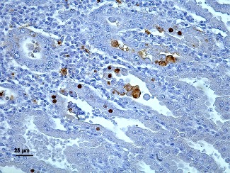
Histopathological examination of the proventriculus showed multifocal necrosis of oxynticopeptic cells together with a moderate intraglandular multifocal lymphocytic infiltrate in seven of the nine studied birds (three, three and one from farms A, B and C, respectively). Moreover, occasional swollen nuclei with marginated chromatin and clear centres resembling intranuclear inclusion bodies were also observed. On the basis of these findings, TVP was diagnosed in all three flocks. Using electron microscopy, several viral particles approximately 60 to 70 nm in size, with icosahedral symmetry and without an envelope, were observed within the cytoplasm of necrotic epithelial cells. Taking into account their location and morphology, these particles were compatible to virus from the Reoviridae or Birnaviridae families. Immunohistochemistry using monoclonal antibody to IBDV protein VPX was performed. Positive staining was observed in the nucleus and/or in the cytoplasm of oxynticopeptic cells in the proventriculus of the seven chickens showing TVP-compatible lesions, highly associated with affected areas. No other tissues, including bursa of Fabricius, gave positive results by immunohistochemistry. On the other hand, PCR on RNA extracted from paraffin-embedded proventriculus using specific primers for the VP2 gene of IBVD gave negative results.
Multifocal necrosis of oxynticopeptic cells together with moderate intraglandular multifocal lymphocytic infiltration in the proventriculus of a chicken. A nucleus of a glandular cell showing nuclear chromatin margination and pale-stained central area is shown (arrow). Haematoxylin and eosin. Bar=25 µm.
Although intranuclear inclusion bodies have not been described in IBD, the most important and known disease of chickens caused by IBDV, the immunohistochemical and ultrastructural results obtained in our studies suggest that the detected IBD-like virus was the aetiologic agent of TVP in the cases presented here. Nuclear changes have been mentioned before in TVP cases, usually described as nuclear chromatin margination and pale-stained central areas. Further studies are needed to elucidate the origin of nuclear changes, as well as to characterise in detail the detected viral agent. Efforts are now focused on amplifying and sequencing the intralesionally detected IBD-like virus to further characterise it.
References
"Infectious bursal disease-like virus in cases of transmissible viral proventriculitis". L. Grau-Roma, A. Marco, J. Martínez, A. Chaves, R. Dolz, N. Majó. Veterinary Record, 2010;167:836 doi:10.1136/vr.c6561