

Apocrine cystomatosis. First description in the pig species
The study by the Veterinary Pathology Diagnostic Service (SDPV) of the UAB and the Slaughterhouse Support Network (SESC) of the IRTA-CReSA for the first time describes apocrine cystomatosis in the porcine species, a disease of the apocrine sweat glands already studied in other animal species such as cats and dogs. Based on three cases in Catalonia, this study states that although the cause of the condition is the same, it has not been possible to determine one single cause in general (local or multifocal cutaneous or systemic) due to the lack of a wider sample analysis. Nonetheless, all signs point to the possibility of a congenital origin.
Skin diseases are relatively common in pig production and are often the cause of seizure of the carcass at the slaughterhouse level. The clinical presentation of this type of pathologies ranges from focal lesions that manifest exclusively at the cutaneous level or as multifocal or generalized lesions of systemic origin, often accompanied by other extracutaneous clinical signs.
Determining the origin of these diseases is complex for veterinary specialists in pigs or slaughterhouse inspectors since there are many causes that produce them and are of different origins. The most common are congenital, nutritional, environmental, infectious diseases of cutaneous or systemic origin and neoplastic causes among others.
Proliferative skin lesions are common in pigs. The age of presentation helps determining the most likely origin, with tumors being the most frequent cause in adult animals. In the case of young animals, the most likely causes are congenital or, to a lesser extent, of infectious origin. However, neoplastic diseases are not as frequent as other causes since animals are sent to slaughter at an early age, before reaching sexual maturity, when tumors develop more frequently. Despite this, cutaneous neoplasms have been described in young pigs, with lymphoma, nephroblastoma and melanoma being the most frequent tumors at these ages. Apocrine cystomatosis, also known as cystic hyperplasia of the apocrine sweat glands, is a rare skin condition of non-tumor origin that, so far, has been described in humans, dogs and, less frequently, in cats but not in pigs or any other species used in livestock.
This work describes for the first time an apocrine cystomatosis in three 6 months old pigs during the post-mortem inspection in different slaughterhouses in Catalonia. The cases were referred through our slaughterhouse support service through SESC. In all three cases the characteristics of the lesion were the same.
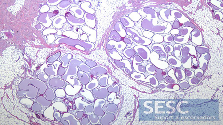
Macroscopically, multiple homogeneous vesicles of less than 1 cm in diameter were observed aligned and filled with transparent liquid distributed throughout the subcutaneous tissue of the skin on the back of the animal. At the histopathological level, it was confirmed that these vesicles consisted of distended sweat glands filled with ecstatic material with no associated inflammatory reaction.
Although the location of the sweat gland is usually the same within the same species, the anatomical location of them varies among the different species of domestic animals. In the three pigs presented, the location of the lesion on the back of the animal seemed to follow this norm but the low casuistry does not confirm this hypothesis.
In veterinary medicine, no consensus has been reached on the cause of apocrine cystomatosis. An accumulation of the material produced by the sweat glands after obstruction of the excretory duct could lead to glandular distention. However, in these cases the expected lesion would be focal not generalized. Another possible explanation for this problem would be the presence of continuous episodes of glandular hyperplasia (which is associated with greater production of material) followed by processes of glandular involution and impossibility of eliminating the material, which leads to its accumulation. Another possible cause, which is also reported, is the presence of benign tumors (adenomas) of the sweat gland. These neoplasms are characterized by growing uncontrollably but progressively maintaining the production capacity. This leads to the accumulation of material and the consequent glandular distention. The diagnosis of adenomas requires the identification of microscopic characteristics indicative of cell proliferation, although this was not confirmed in the three cases presented. Finally, the absence of inflammatory cells or infectious agents associated with the glands makes it possible to rule out the infectious cause as responsible for the problem.
A congenital origin cannot be ruled out, but neither can it be confirmed since this lesion is not observable in a live animal. However, the absence of normal apocrine glands in the sections evaluated, the lack of obvious dilation or obstruction of the glandular ducts and / or inflammation or other alterations in the surrounding subcutaneous tissue may be elements that point to a congenital origin.
Àrea de Sanitat Animal
Universitat Autònoma de Barcelona
Institut de Recera i Tecnologia Agroalimentàries - Centre de Recerca en Sanitat animal (IRTA-CReSA)
References
López-Figueroa, C., Domingo, M., Martí, B., Vidal, E., Segalés, J. Cutaneous apocrine cystomatosis in three slaughter-aged pigs. J Vet Diagn Invest. 2020;32(1):159‐161. doi:10.1177/1040638719894553