Porcine circovirosis, pathogenesis and ultrastructural studies
This PhD thesis is an important step in the fight against post-weaning multisystemic wasting syndrome. The disease, in which the porcine circovirus type 2 (PCV2) is the causative agent, produces growth delay and death during the first months of life of the piglet. This innovative research is the first morphogenetic study of PCV2 in cell cultures and provides ultrastructural descriptions of the affected tissues and reveals the relationship of the disease with mitochondries.
Post-weaning multisystemic wasting syndrome (PMWS) is one of the most significant porcine diseases worldwide. This condition is clinically is characterized by growth retardation, weight loss, labored breathing, fever, diarrhea and death in nursery and/or fattening pigs. The essential causative agent is porcine circovirus type 2 (PCV2), the smallest virus known to infect animals. Lymphocyte depletion and infiltration of histiocytes and/or multinucleate giant cells in lymphoid tissues are the hallmark histological features that define the disease; in some cases, intracytoplasmic and intranuclear viral-like inclusions can be found macrophages. Ultrastructurally, PCV2 has been described as a non-enveloped virus of 12-23 nm in diameter.
Several studies have described different biological properties of the virus. It is known that attachment and internalization of PCV2 is accomplished by clathrin-mediated endocytosis and the whole replication cycle takes around 24-36 hours. One of the most classical approaches to elucidate the viral cycle in different cell types includes electron microscopy studies. However, at the initiation of this thesis, little knowledge was available on the subcellular localization of PCV2 in infected cells in vitro as well as in tissues of pigs naturally affected by PMWS. Therefore, the main objective of this doctoral work was to study the ultrastructural alterations associated to PCV2 infection and determine the localization of virus at the subcellular level, describing the morphogenesis of PCV2 on culture cells and assessing ultrastructural findings related with PCV2 virus-like particles (VLPs) in lymph nodes of PMWS affected pigs.
In the first study of the thesis (Chapter 3), the complete morphogenesis of PCV2 in a clone of the lymphoblastoid L35 cell line (L35 cells) was described for the first time using electron microscopy techniques. Cells were infected with PCV2 using a multiplicity of infection of 10 and examined at different time-points post-infection (0, 6, 12, 24, 48, 60 and 72 hpi). A first cytoplasmic phase was described after virus internalization by endocytosis, in which PCV2 particles were initially found aggregated as intracytoplasmic inclusion bodies (ICIs). Subsequently, PCV2 was closely associated with mitochondria. Afterwards, viral factories (VFs) were shown in the cell nucleus. Virus assembly and encapsidation occurred with the apparent participation of the nuclear membrane. Immature virions left the nucleus and formed ICIs in a second cytoplasmic phase. Organelle alterations were associated with the PCV2 infection as assessed by means of immunogold labeling. The ICIs were gold-labeled but not the intranuclear inclusion bodies (INIs). Obtained results suggested that at the end of the replication cycle (between 24 and 48 hours), PCV2 was released by two different ways: by budding of mature virion clusters or by lysis of apoptotic/death cells.
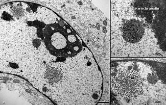
For the second and third studies (Chapters 4 and 5), samples of lymph nodes were taken from PMWS affected pigs and healthy control animals. Significant ultrastructural alterations were noted mainly in histiocytes infiltrating lymphoid tissues from disease animals only. Severe swelling and proliferation of mitochondria, and proliferation and dilation of rough endoplasmic reticulum and Golgi complex were the most significant ultrastructural findings. A significant number of ICIs were observed in close proximity with those damaged organelles. Small and large ICIs were surrounded by double membrane, with a granular appearance or containing paracrystalline arrays. Few numbers of histiocytes had also INIs, which were non-membrane bounded. Icosahedral VLPs were 8-17 nm in diameter and were labeled by means of a PCV2 monoclonal antibody. The VFs were often found next to mitochondria with severe swelling. These mitochondria were associated with PCV2-labeled VFs. As expected, lymphocyte depletion was a striking finding of lymph nodes from PMWS affected pigs. Co-localization studies with confocal microscopy and double immunolabeling with TEM indicated a close relationship between VFs and mitochondria, suggesting that these organelles may play an important role in the replication of PCV2. The present findings further support the hypothesis that virus replicates within the histiocytes of lymph nodes.
In summary, the present thesis includes the first morphogenesis study of PCV2 on cell culture as well as the most detailed ultrastructural descriptions of lymph nodes from PMWS affected pigs. Obtained results allowed to complement existing works on the PCV2 replication cycle in cell culture and to gain new insights on the pathogenesis of PMWS. The description of the intimate association between PCV2 and mitochondria is one of the major contributions of the present thesis.
References
"Ultrastructural studies on Porcine circovirus type 2 (PCV2) infection". PhD thesis defended by Carolina Rodríguez Cariño, July 13, 2010. Director: Dr. Joaquim Segalés i Coma.