

Sensory Stimuli Improves Brain Damage in Mouse Models of Preterm Birth
Perinatal brain injuries hinder neurological capabilities throughout life, causing anything from fine motor problems to severe cognitive limitations. At the same time, therapy treatments currently available are very limited. That is why other types of interventions to help counter these effects are being explored.
In this context, a new longitudinal study demonstrates for the first time in a murine model of premature infants with brain injury caused by hypoxia and ischemia that the behavioral, cognitive and emotional effects in their childhood and adulthood depend on gender, age and task. However, these effects can be modulated by neonatal tactile and proprioceptive stimulation, especially in males.
The study was published in a special edition of the journal Frontiers in Behavioral Neuroscience, edited by Professor Rosario Montirosso from the Eugenio Medea Research Institute of the Italian Ministry for Health. The special edition is dedicated to an international collection of new evidences on humans and animals regarding Risk and Protection Factors Associated With Adversity in Early Stages and Infant Development. The research publication has been reviewed by the experts Dr. Michael A. Van Der Kooij from the Johannes Gutenberg University Mainz, Germany, and Dr. Sharon Casavent, from the University of Connecticut, USA.
The importance of this work lies in that the immature brain of preterm infants, equivalent to that of mice when born, is at a larger risk for hypoxic-ischemic damage, and male newborns are more susceptible and respond worse to protective and therapeutic interventions.
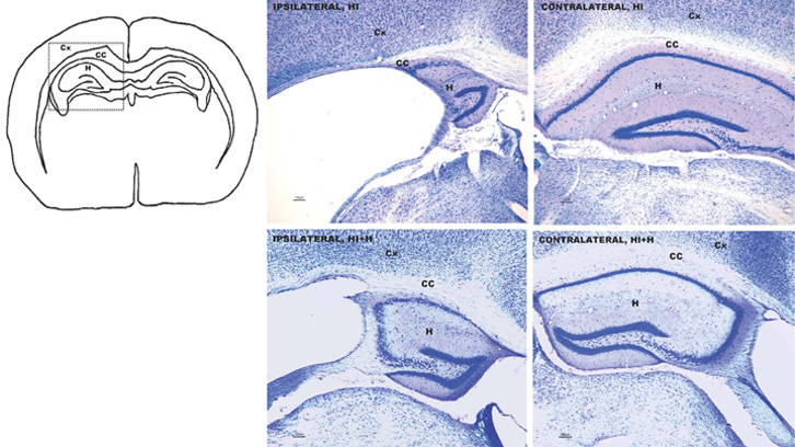
Although the hypoxic and ischemic lesions induced similar brain damage in males and females, the effects were different depending on gender, age and specific task evaluated. During the infant stage, the damage affects balance, particularly among females, and prehension (organ or appendix’s ability to take or catch something) in males, but both aspects improve as they grow and only reflexes remain damaged. Male mice showed to have infantile hyperactivity, which normalized as they became adults. In contrast, the anxiety and emotional traits of these injuries lasted throughout their lives. Both genders showed poorer learning processes at short and long terms, but there was more damage to memory among the males. The functional evaluations were correlated with the degree of severity of the affected brain areas: hippocampus, caudate/putamen, thalamus, neocortex and corpus callosum.
Sensory stimulation, applied in mice from before the injury occurred until the final stages of infancy, exercised a remarkable neurological protection, especially in males. The neurological protection in both genders was correlated to the improvement of functional capacities, reflexes, and an improvement in memory results.
As a whole, the study shows the different neuronal substrates needed to satisfy functional demands and points to the most resilient neuroanatomical targets to repair these functions through postnatal stimulation.
Despite the obvious differences between rodents and humans, the study shows the complex relationship between different regions of the brain, risk factors, vulnerability and resilience, and all dependent on gender and age. It also provides new data on behavioral neuroscience within the field of neonatology and the area of pediatric functional rehabilitation, defining a translational scenario in which to study the underlying mechanisms of the functional and neuropathological correlates found.
The origins of the study conducted by researchers of the INc-UAB includes the collaboration of Dr Lydia Giménez-Llort, Director of the Medical Psychology Unit of the Department of Psychiatry and Forensic Medicine of the UAB, and Dr Laia Acarin, researcher from the Medical Histology Unit of the Department of Cell Biology, Physiology and Immunology. It was conducted under the framework of the La Marató* de TV3 project committed to brain injuries, in a common interest to join forces in solving severe neurological problems arising in infancy and finding mechanisms with which to improve the functional capacities throughout their lifetime.
Institut de Neurociències (INc-UAB)
References
A Muntsant-Soria, K Shrivastava, M Recansens, L Giménez-Llort. (2019). Severe Perinatal Hypoxic-Ischemic Brain Injury Induces Long-term Sensorimotor Deficits, Anxiety-like Behaviors and Cognitive Impairment in a Sex-, Age- and Task-Selective Manner in C57BL76 Mice but Can Be Modulated by Neonatal Handling. Frontiers in Behavioral Neuroscience, 13:7, 1-19. DOI: 10.3389/fnbeh.2019.00007/full.