Novel MRI Contrast Agents for Brain Tumor Diagnosis
Researchers from the UAB and the Institut Català de Nanociència I Nanotecnologia (ICN2) have developed a nanoscale coordination polymer particle (NCP) built using paramagnetic iron(III) metal ions able to exhibit T1 and T2 high contrast potential for the magnetic resonance image (MRI) in vivo, allowing simultaneous recording of positive and negative contrast images in a very short period of time. This work opens new possibilities in constructing easily biodegradable and nontoxic nanoparticles with improved T1/T2 dual-mode MRI contrast enhancement for tumour diagnosis.
One of the most used methods for the diagnosis and the follow up treatment of brain tumours is MRI (magnetic resonance image). Contrast agents (CAs) are sometimes needed to better highlight tumours, being the most used the paramagnetic Gadolinium-based T1 CAs, but their use is limited by renal toxicity, rapid clearance, and need for repeated administration, as well as inaccuracies in delineating tumor margins. Therefore the development of a non-toxic contrast agent that improves the tumor imaging and that persists in the tumor and could assist in image-guided radiotherapy, as well as in the tumor resection and monitoring of response early during the course of therapy could be of interest.
A study led by Dr. Fernando Novio, researcher at the Catalan Institute of Nanoscience and Nanotechnology (ICN2) and associated professor at the Chemistry Department of the Autonomous University of Barcelona (UAB), and Dr. Julia Lorenzo, researcher at the Institute of Biotechnology and Biomedicine (IBB) and professor at the Biochemistry and Molecular Biology Department (UAB), has developed an iron-based nanoscale coordination polymer (Fe-NCP) that exhibit high biocompatibility, low toxicity and dual-mode T1 (bright images) and T2 (dark images) MRI contrast. The dual-mode CA capability was evaluated in vivo using a preclinical model of murine glioblastoma. The in vivo MRI of Fe-NCPs show T1 and T2 high contrast potential, allowing simultaneous recording of positive and negative contrast images of the brain tumors in a very short period of time while being safer for the mouse. The comparative study with commercial CAs suggest that these nanoplatforms are promising candidates for the development of dual-mode MRI CAs with enhanced contrast and overcoming the main drawbacks of commercial and other reported CAs.
This work was possible thanks to the collaboration with Dr. Carles Arús and Dra Ana Paula Candiota from the Grup d´Aplicacions Biomèdiques de la Ressonància Magnètica Nuclear GABRMN (UAB).
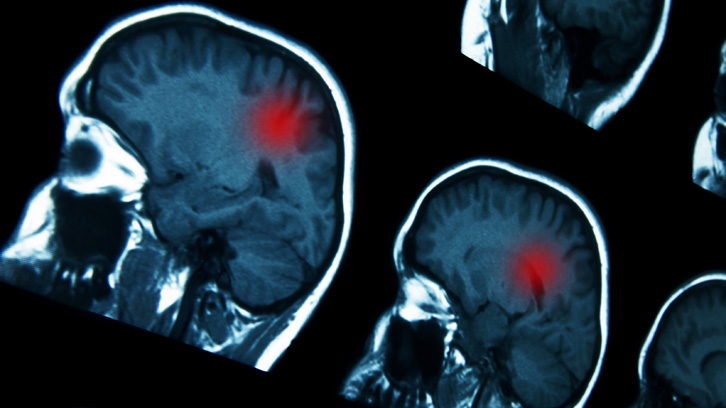
Figure 1. a) Schematic of the Fe-NCPs; b) Scanning electron microscopy image of the resulting nanoparticles; c) MRI contrast behaviour of Fe-NCPs in solution (phantoms); d) merge imaging of in vivo brain tumor combining T1 and T2 images generated by Fe-NCPs.
Julia Lorenzo Rivera 1 i Fernando Novio 2
1Institut de Biotecnología y de Biomedicina (IBB) y Biochemistry and Molecular Biology Departamento de Bioquímica
Suárez-García S, Arias-Ramos N, Frias C, Candiota AP, Arús C, Lorenzo J*, Ruiz-Molina D, Novio F*. (2018). Dual T1/ T2 Nanoscale Coordination Polymers as Novel Contrast Agents for MRI: A Preclinical Study for Brain Tumor. ACS Appl Mater Interfaces. Nov 14;10(45):38819-38832. DOI: 10.1021/acsami.8b15594.