

Neurogenesis in adult ventricular-subventricular area as the origin of glioblastoma multiforme
The existence of neurogenesis in the central nervous system of adult mammals was first demonstrated in the 1960s thanks to the investigations of Dr. Joseph Altman.
It is now widely accepted that there are two neurogenic areas, the first located in the subventricular region of the deat gyrus and the other in the ventricular-subventricular region.
In both areas, neural stem cells stem cells with proliferative capacity persist in mature individuals. These generate neurons, astrocytes and oligodendrocytes through a complex network made up of cellular precursors and their descendants.
It is now accepted that neural stem cells have their origin in embryonic radial glial cells and therefore, neurogenesis in adults is interpreted as an extension of the embryonic.
Glioblastoma multiforme is a type of cerebral tumor of high aggressiveness and with little expectations of cure, because it is refractory to the chemical and radiological therapies.
This resistance seems due to the marked cellular heterogeneity that these tumors show, the origin of these cellular populations is not known with exactitude.
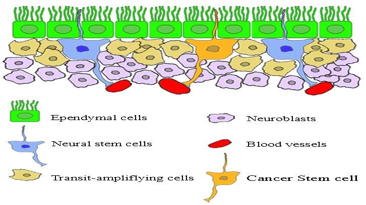
The present study has examined the relationship between ventricular-subventricular area stem cells, the onset and development of glioblastoma multiforme. The presence in the neurogenic region of some stem cells characterized by being mitotically active and at the same time resistant to chemotherapeutic agents, makes these cells candidates to explain the origin of glioblastoma multiforme.
Specifically, they can lead to the formation of so-called cancer stem cells. However, both neural stem cells and cancer stem cells share morphological and molecular characteristics.
As an example, the expression of Nestinar, glial fibrillar acid protein and β-tubulin III, among others, has led to hypothesize that the cancer stem cells have an embryonic origin.
In a glioblastoma multiforme, the cancer stem cells represent only a small cell fraction with proliferative capacity. However, these cells show several characteristics, among them the ability to self-perpetuate and to originate different cellular populations, also with mitotic capacity.
The latter contribute significantly to the size of the tumor, despite some contradictions, the CD133 or prominin-1 glycoprotein has become a marker of the cancer stem cells.
A very interesting histological feature of the cancer stem cells is the fact that they are located in those parts of the tumor where the vascularization is weak, irregular and at the same time chaotic.
There are studies that show a marked relationship between the degree of vascularization of a glioblastoma multiforme and its aggressiveness. In addition hypoxia, more aggressiveness and less response to treatments.
Finally indicate that different lines of research indicate that hypoxia contributes significantly to the development and expansion of cancer stem cells, at the same time would form a microenvironment that would allow them to survive the therapies used so far.
Faculty of Biosciences
Universitat Autònoma de Barcelona
References
Claudia Capdevila, Lucía Rodríguez-Vázquez and Joaquim Martí-Clúa. Glioblastoma Multiforme and Adult Neurogenesis in the Ventricular-Subventricular Zone: a Review, J. Cell. Physiol. 232: 1596-1601 (2017).