

Hydroxyurea injection leads to apoptotic and autophagic cellular events in the cerebellar external granular layer
The cerebellar external granular layer is a temporary structure involved in the generation of granular neurons. This germinal matrix presents neuroblast with a high proliferative activity. This current study analyzes, in rats, the effect of hydroxyurea exposition on the neuronal progenitors of the aforementioned structure. Our results allow to know the changes that occur in the cerebellar architecture, as well as the cellular responses produced as a consequence of the administration of cytostatic agents.
The histological study of the development of the cerebellum shows that the neural populations that constitute this region of the central nervous system are generated, migrate and finally settle in their places of destination following a marked temporal and spatial regularity. These peculiarities make the cerebellum an ideal model for knowing how certain pharmacological treatments can alter its normal development and therefore the spatial arrangement of its cellular components. Our study aimed to analyze the effect of an inhibitor of the ribonucleotide reductase enzyme, called hydroxyurea, on the development of the cerebellar external granular layer. This germinal matrix is responsible for originating the granule cells. In particular, it has determined whether this drug causes apoptosis and autophagy in the neuroblasts of the external granular layer. Hydroxyurea is a drug used for the treatment of various types of cancer and myeloproliferative diseases. It is included in the World Health Organization’s list of essential medicines.
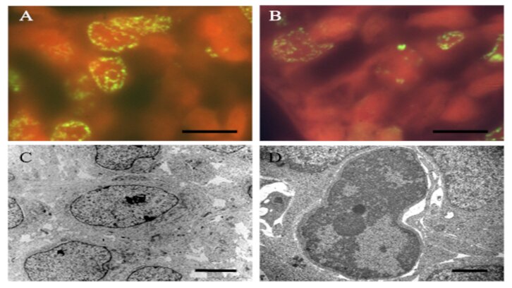
To do this study, 9-day-old rats were injected with a single dose of saline or 2mg / kg of hydroxyurea (intraperitoneal), and sacrificed at appropriate times ranging from 10 to 60 h. The use of cytochemical, immunohistochemical, and electron microscopy techniques indicate that the administration of hydroxyurea leads to the activation of apoptotic cellular events in the external granular layer. The resulting data denoted that the administration of hydroxyurea leads to the activation of apoptotic cellular events that began to increase 10 h after hydroxyurea exposure, peaked at 30 h, and decrease thereafter. It also showed that apoptosis was followed by autophagy activation. Interestingly, LC3B and p62/SQSTM1-stained cells, as well as mitotic cells, started to appear 20 h after the HU-injection and their counts increased until 40 h. Afterwards, the values remained stable.
The current results reveal that the administration of HU first induces the apoptotic cell death and after that, cellular profiles that meet criteria for autophagy start to appear. These effects produce important alterations in the cytoarchitecture of the cerebral cortex. These conditions persist into adulthood. We propose that the treatment with hydroxyurea induces an autophagic response that counteracts the apoptosis produced by this drug. In other words, neuroblasts in the external granular layer may activate an autophagic response that serves as a survival mechanism against HU. In addition, these results provide a clue for studying the mechanism of chemoresistance triggered by proliferating cells exposed to anticancer agents.
Universitat Autònoma de Barcelona (UAB)
Cytology and Histology Unit
Department of Cell Biology, Physiology and Immunology
Faculty of Biosciences
Institute of Neurosciences
References
Vanessa Molina, Lucía Rodríguez-Vázquez and Joaquin Martí. Patterns of Apoptosis and Autophagy Activation After Hydroxyurea Exposure in the Rat Cerebellar External Granular Layer: an Immunoperoxidase and Ultrastructural Analysis. Neurotoxicity Research (2020) 37:93-99. https://doi.org/10.1007/s12640-019-00094-y