

Alba synchrotron may help in the fight against cancer
In the last 30 years many groups have being carrying out experiments and trials of new techniques in image and radiotherapy of cancer using synchrotron X rays.
Synchrotron radiation (SR), combines extremely high photon beam intensity, small apparent source size, high collimation, tunability, and a continuous energy spectrum (from the far infrared region up to hard X-rays). These characteristics make the SR a unique and useful tool for biomedical imaging and therapy. Some of these methods show themselves great promise for the treatment of cancer.
Imaging Techniques
Conventional radiography produces images through the differential absorption of X-rays through the tissues, whereas synchrotron-based imaging techniques can also produce high-resolution images using differences in the refraction and scatter of X-rays.
Analyzer-based Imaging(ABI)
ABI is a new x-ray imaging technique that uses monochromatic radiation to produce a refraction map of the object. The technique is based in the use of a perfect crystal between the sample and the detector. This crystal, acts as an angular slit, and transforms the angular deviation caused by refraction in the object into intensity variations at the detector.
Propagation based phase-contrast Imaging (PBI)
PBI technique is based of the interference pattern produced by the transmitted waves of the x-ray beam. The interference pattern shape depends on the distance to the detector, and several patterns can be recorded at different distances, to reconstruct the phase-shift across the sample. If the detector is placed immediately after the sample, a conventional absorption image will be obtained. With the detector placed very far, one will observe a fringe pattern as different components of the diffracted beam interfere with each other. This regime corresponds to Fraunhofer -or Fairfield- diffraction. The interference pattern contains useful phase information, but extracting it is an ongoing computational and physics challenge. If the detector is placed at an intermediate distance, one gets Fresnel or near-field diffraction pattern. In this case, a combination of absorption and diffraction effects is found, as Fresnel fringes. These fringes improve the edge visibility, allowing us to see the density contours in the sample, which cannot be seen through simple absorption contrast.
K-edge subtraction(KES)
KES consists of measuring the attenuation of two x-ray beams whose energies are slightly above and below the K-edge of an element (contrast). This contrast agent it is chosen to have an absorption K-edge in the range on energies of the image, and with an appropriate attenuation coefficient. Since most elements and compounds in the body change very little in a narrow energy range, but not the selected element, two images at different energies can be subtracted, and hence the image of the physical distribution of the contrast agent is obtained. The resultant image maximizes the contrast agent while minimizing signals arising from other parts of the tissue.
Scattering techniques
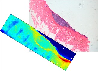
Radiation across the matter may be scattered as-well as refracted or absorbed. Scattered photons bear information about the molecular structures of the materials. Small-angle x-ray scattering (SAXS) is a technique to study the structures of materials at scales that range from molecules to almost cellular size structures. It has been demonstrated that the supra-molecular structure of collagen it is different in pathological tissues than those in normal benign tissues. SAXS allows study these differences and correlated with pathological state of connective tissues, as in the case of breast cancer or melanoma.
Radiotherapy Techniques
Theradiotherapy technologies from Marie Curie to date, has always focused its development, trying maximizing the deposited dose in pathological tissue and minimizing the damage on healthy surrounding and ones.High intensity and collimation of synchrotron X-rays will offer novel methods of radiation therapy.Synchrotron radiotherapy makes possible very precise tumour targeting with an extremely high dose rate up to thousands of grays in a few milliseconds. Different strategies are followed on synchrotron radiotherapy experimental treatments; polychromatic but spatial fractionated radiation (MRT and MBRT) or monochromatic ones (SSRT).
Microbeam Radiation Therapy (MRT)
This technique is based on the idea that radiation damage that induce tissue necrosis can be considerably reduced by spatial micro fractionation of the absorbed dose. MRT beam is split by collimators into many parallel and planar beams in the 50 – 150 keV energy range. The thickness of each microbeam is 20–50 μm with a separation of 100–200 μm
Different experiments have shown that microplanar beams yields a larger therapeutic index than does broad beam irradiation for the intra-cerebral rat 9L gliosarcoma (9L GS)[22], for the murine mammary carcinoma (EMT-6)[23] and for the aggressive murine squamous cell carcinoma (SCCVII). Moreover, adult brains of rats, duck embryos and piglets have shown to display an unusually high resistance to damage when they are irradiated by single-exposure unidirectional microplanar beams, tolerating doses up to 10-fold larger than that of broad-beam irradiation.
Minibeam Radiation Therapy (MBRT)
In the MBRT, the beam thickness ranges from 500 to 700 micrometers with a separation between two adjacent minibeams of the same value. While such thin microbeams can only be produced by synchrotron sources and have other practical limitations to clinical implementation, MBRT could conceivably be implemented with high power orthovoltage X-ray tubes. Our group is studying the different effects in vitro that induced the MBRT over F98 rat glioma cells. Data obtained led to detect more effectiveness of minibeams, These experiments also show that the side effects in the healthy tissues to be much reduced in MBRT.
Sterotactic synchrotron radiation therapy(SSRT) and Photon Activation Therapy (PAT)
PAT and SSRT are two-step techniques where firstly, a vehicle transports and delivers high Z atoms near or attached to DNA nucleus of tumour cells.A subsequent synchrotron irradiation with energy slightly above the K-edge absorption of these target atoms leads the emission of Auger electrons and photoelectrons which both deposit their energy at the immediate vicinity.
The utilization of KeV energy range carries with it less side effects compared with the MeV conventional therapeutic radiation.
All these in vitro experiments could be extended to another radioresistant tumour cells in order to transfer the methodology or some future developed technologies to the hospital applications.
No later than one year 7 beam stations will start to work in Alba synchrotron, none of them will be devoted for biomedical applications, as difference of synchrotrons of Canada, Australia both also new synchrotrons with similar technology than Alba. More than 10 hospitals and other more than 10 health research centers that exist near Alba (around 20 km.). This suggests that if there were a consortium research center in imaging and radiotherapy in the technological park of Alba, linked with the synchrotron would become a center of excellence in this field and will induce the construction of a biomedical beam line. This project will set a new development and transference laboratory for a new technologies and applications in imaging and radiotherapy