Crucial role of mRNA in cellular tumor control and response revealed
A pioneering UAB study reveals the fundamental role of secondary structures of encoding mRNA in the DNA damage detection machinery, which directs and stabilizes vital proteins in the response to genetic damage, such as p53. The findings suggest new therapeutic and diagnostic implications, opening doors to understanding the molecular mechanisms underlying diseases such as cancer.
DNA, the molecule that contains all the genetic information inside cells, is constantly exposed to several threats that can damage its integrity. To counteract this damage, cells have evolved a sophisticated surveillance and repair system known as the DNA damage detection machinery. This system, composed of a set of proteins (such as KRAS, IGF-1 and p53), deploys an intricate molecular choreography to detect and correct damage that arises in the DNA structure, thus ensuring genetic stability and proper cellular functioning. Therefore, these proteins involved in the DNA damage detection machinery are of particular interest in the world of cell biology because of their crucial role in the preservation of genetic integrity and tumor control.
Our study, published in the Molecular Cancer journal, is the first demonstration of how a secondary structure of coding mRNA programs the DNA damage detection machinery to direct downstream factors (proteins, enzymes, or genes that carry out various cellular functions, such as division, differentiation or even death), leading to stabilization and activation of the encoded protein.
Recent evidence points towards a groundbreaking mechanism whereby mRNA secondary structures (the conformation in which the nucleotide sequence of the molecule takes in space) alter the expression levels of the encoded proteins. These RNA structures can be altered by synonymous mutations (SMs: mutations that do not alter the protein but alter the RNA transcript). Even though SMs have a significant occurrence of 6–8% in driver mutated genes in cancer and accumulating evidence points towards functional implications of SMs, studies resolving the underlying cellular mechanism are challenging and there is a need for more results. In this research, we have used the tumor suppressor called "p53" as a model to investigate the role of secondary structures in the activation of the encoded protein.
The p53 protein is a transcription factor (a protein that regulates gene expression) that is found mutated in more than 50% of cancers in humans and plays a pivotal role in controlling the behavior of the cell in response to cell stress. One of these behaviors is to signal the cell towards cell apoptosis (death), thus preventing the spread of cancer. The activation of p53 is regulated in the cell by the protein ATM kinase and forms an essential part of the DNA damage response (DDR) and genotoxic stress. Since under normal conditions there is no DNA damage and the cells need to grow and divide, p53 is maintained a low levels (inactive). Another protein, the MDM2 E3 ubiquitin ligase, interacts with p53 protein and mediates a modification on p53 that signals the cell to degrade it. However, during the genotoxic stress, the cell suffers from DNA damage and needs p53 to stop its erroneous division. As such, the ATM kinase is in turn activated and modifies (phosphorylates) the MDM2. This modification allows MDM2 to instead bind the p53 mRNA and not the p53 protein. This interaction mediates the formation of a complex of MDM2 with p53 mRNA and ribosomal proteins and mediates its translocation to the cytoplasm, at the p53 polysome, where ATM activates p53 towards the DNA damage response.
We advanced our previous findings by focusing on the upstream DNA Damage Sensing machinery involving ATM and the MRN machinery. Specifically, we employed a combination of in vitro (biochemical) assays, as well as the Proximity Ligation Assay in human cell lines, to reveal the dynamic sub-cellular localization of these interactions during genotoxic stress, and the phosphorylation events on ATM that act synergistically to control the interaction and its trafficking to the cytoplasm (cellular studies). Thus, we show for the first time that ATM protein binds the p53 mRNA and this event leads to the MDM2-dependent activation of p53.
Therefore, this study advances pioneer concepts of functional roles of the mRNA, which are potentially broadly employed in cell signaling, and reveals how p53 synonymous mutations can take up roles as therapeutic or diagnostic targets in precision medicine.
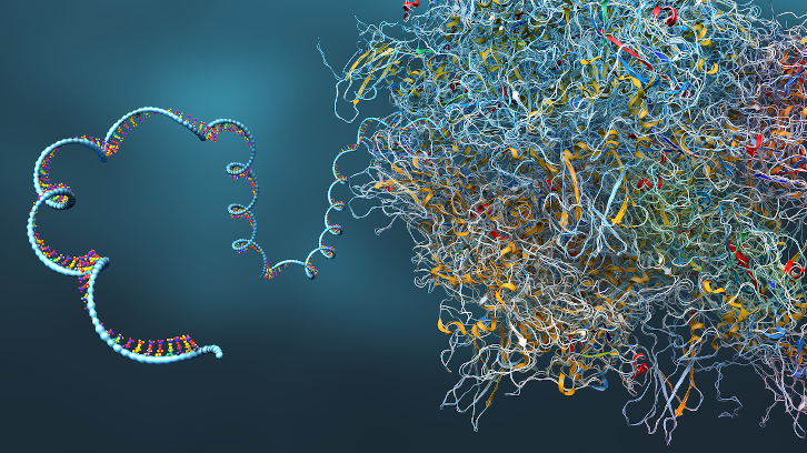
Model describing how the ATM exchanges the p53 mRNA substrate with MDM2, ultimately leading to the phosphorylation of the nascent p53 peptide (p53-Ser-15) that activates p53 towards the DNA damage response.
Konstantinos Karakostis
Institute of Biotechnology and Biomedicine
Universitat Autònoma de Barcelona
References
Karakostis et al. Molecular Cancer (2024) 23:21 https://doi.org/10.1186/s12943-024-01933-z