Synchrotron light to treat brain tumours: divide and win
One of these research lines studies fragmentation in a synchrotron of X-ray beams used in radiotherapy. Smaller, subdivided beams converge in the tumour but produce less damage to the tissues they have gone through. UAB researchers have compared this technique with conventional radiotherapy in rat gliomas, and discovered that the division of beams can more effectively attack the tumour while minimising damage to surrounding tissues.
Gliomas are among the most frequent primary brain tumors in adults, with an incidence of approximately 5/100,000 among the general population, treatment of high-grade gliomas is only palliative. A radical radiotherapy treatment of radioresistant tumors would require the development of new techniques allowing sparing the sensitive surrounding normal tissue.
Since 1990s synchrotron radiation has become one of the most valuable tools in experimental radiotherapy in the quest for a radical treatment for gliomas. Synchrotron sources are ideal for spatially fractionated techniques such as Microbeam Radiation Therapy (MRT) and Minibeam Radiation Therapy (MBRT), currently under development at the European Synchrotron Radiation Facility -ESRF- in Grenoble, France. The beam width is 25 μm in MRT, whereas in MBRT beams of 700 μm width are employed. The energy spectrum employed ranges from 50 to 500 keV, and with a mean energy at around 100 keV.
The sparing effect observed in MRT, supporting a potential application of minibeams to treat tumors with minimal damage to the surrounding healthy tissues.
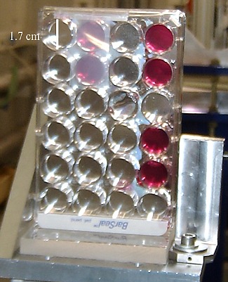
Our groupcarries out vitro studies, using the MBRT against F98 glioma rat cells. Plates were irradiated on the position shown in Figure 1 and increasing doses of 6,12,18,24, 30 and 40Gy.
Figure 2A
Figure 2B
F98 glioma cells were irradiated with spatially fractionated and seamless synchrotron x-rays. The percentage of each cell population (alive, early apoptotic and dead cells, where either late apoptotic as necrotic cells are included) was assessed by flow cytometry 48 hours after irradiation, whereas the metabolic activity of surviving cells was analyzed on days 3, 4, and 9 post-irradiation by using Q-Blue test. See figures 2A, 2B, and 3.
Figure 3
Similar cell survival ratio and cell recovery were observed between cells irradiated with MBRT or BB. However, a significant increase in the number of early apoptotic cells was found 48 hours after the minibeam radiation in comparison with the seamless mode.
The endpoint (or threshold dose from which an important enhancement in the effectiveness of both radiation treatments is achieved) obtained by flow cytometry could be established just before 12 Gy.
The two irradiation schemes, whilst the endpoints assessed by the QBlue reagent, taking into account the cell recovery, were set around 18 Gy in both cases.
MBRT might offer probably a remarkable healthy tissue sparing in comparison to BB, as already observed in some first experiments.
References
"Survival Analysis of F98 Glioma Rat Cells Following Minibeam or Broad-Beam Synchrotron Radiation Therapy" Silvia Gil, Sukhéna Sarun, Albert Biete, Yolanda Prezado and Manel Sabés. Radiation Oncology 2011, 6:37 doi:10.1186/1748-717X-6-37.