Teat skin colonization with contagious mastitis pathogens increase the risk of bovine intrammary infections
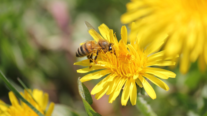
Intramammary infections (IMI) with Staphylococcus aureus and Streptococcus agalactiae are usually associated with subclinical infections that reduce milk quality and quantity. The main transmission route of these contagious mastitis pathogens is through transfer from cow to cow during milking. The teat skin might therefore serve as a reservoir of pathogens that enter the udder through the teat canal and cause IMI. It is generally agreed that Staph. aureus can be isolated from the teat skin and other extramammary body sites, and teat skin colonization with Staph. aureus has subsequently been epidemiologically associated with Staph. aureus IMI.
To avoid these infections, a more intense hygiene during the milking is recommended even though there is no direct cause related to it. That is why it is important to know the role of these reservoirs. In fact, the role of the teat skin as a source of Staph. aureus has not been investigated in herds with automatic milking systems (AMS), where milking hygiene and teat cleaning may differ from the traditional milking systems. Lactating dairy cows can be milked several times a day in AMS without being in contact with human hands, and up to 60 cows can be milked with the same milking unit. These factors are likely to affect teat skin colonization and the transmission of contagious mastitis pathogens. To the best of our knowledge, the association between Strep. agalactiae on teat skin and in milk has not yet been studied. Strept. agalactiae was isolated from teat skin and other areas on cows and in cowsheds. However, Strep. agalactiae was still considered an obligate intramammary pathogen until an environmental reservoir was recently suggested, as Strep. agalactiae was isolated from, for example, water troughs, milking robots, and stalls, and the rectum and vagina of the cows.
Although bacterial culture has mostly been used to study the above-mentioned aspects, PCR has generally been used more frequently in recent years, particularly in European countries. The PCR assay may have a higher analytical sensitivity, and the potential to detect a broader range of bacteria simultaneously without additional diagnostic efforts. However, the PCR assay may detect nonviable bacteria, which have no important role in transmission. A serious implication of nonviable bacteria is that false-positive reactions and subsequently misdiagnosis. On the other hand, viable bacteria only are detected by bacterial culture. However, nonviable bacteria may also be an expression of past exposure, where the bacteria have been killed by teat disinfectants or other circumstances. As such, bacterial culture may be considered more specific for some interpretations. Therefore, the use of both methods may provide information on slightly different aspects of the pathogens in the udder and the surroundings.
From a prophylactic point of view, knowledge of pathogen reservoirs is crucial in the management of Staph. aureus and Strep. agalactiae transmission to prevent IMI. Additionally, in large dairy herds with AMS, controlling transmission related to milking is fundamental in reducing the number of new infections with contagious mastitis pathogens.
We carried out this study to investigate the association between colonization of the teat skin and IMI with Staph. aureus or Strep. agalactiae in the same quarter in dairy herds with AMS.
Milk and teat skin samples were collected from 300 dairy cows with subclinical mastitis from 8 herds and both techniques, PCR and bacterial colonies, were used to detect the different pathogens in the udders and surroundings.
Our findings showed that bacterial culture detected Staph. aureus in 93 (8.1%) of the milk samples and 75 (6.6%) of the teat skin samples. Of these, 15 (1.3%) quarters were positive in both the teat skin and milk samples. Strept. agalactiae was cultured in 84 (7.4%) of the milk samples and 4 (0.35%) of the teat skin samples. Of these, 3 (0.26%) quarters were positive in both the teat skin and milk samples.
The PCR detected Staph. aureus in 29 (10%) of the milk samples and 45 (16%) of the teat skin samples. Of these, 2 (0.7%) quarters were positive in both the teat skin and milk samples. Strept. agalactiae was detected in 40 (14%) of the milk samples and 51 (18%) of the teat skin samples. Of these, 16 (5.6%) quarters were positive in both the teat skin and milk samples.
Based on bacterial culture results, teat skin colonization with Staph. aureus resulted in 7.8 (95% confidence interval: 2.9; 20.6) times higher odds of Staph. aureus IMI, whereas herd was observed as a major confounder. However, results from the PCR analyses did not support this association. Streptococcus agalactiae was isolated from the teat skin with both PCR and bacterial culture, but the number of positive teat skin samples detected by culture was too low to proceed with further analysis. Based on the PCR results, Strep. agalactiae on teat skin resulted in 3.8 (1.4; 10.1) times higher odds of Strep. agalactiae IMI.
We concluded that the presence of Staph. aureus and Strep. agalactiae on teat skin increase the risk of IMI with the same pathogens. Focus on the teat skin hygiene is still recommendable; however, no causal relation can be established.
Universitat Autònoma de Barcelona
References
Svennesen L, Nielsen SS, Mahmmod YS, Krömker V, Pedersen K, Klaas IC. (2019). Association between teat skin colonization and intramammary infection with Staphylococcus aureus and Streptococcus agalactiae in herds with automàtic milking systems. J Dairy Sci, Jan;102(1):629-639. DOI: 10.3168/jds.2018-15330.