New structural model for the metaphase chromosome based on thin plates
In a previous work of our laboratory we observed plates formed by several layers surrounding human chromosomes partially denatured in the presence of ion concentrations approaching those found in metaphase. This unexpected observation has led us to perform an extensive investigation about these laminar structures. Our transmission electron microscopy (TEM) results show that the plates are also observed in the metaphase chromosomes of chicken, a species with a large evolutionary distance from humans. We have observed that a simple dilution of chromosomes with a hyposmotic solution can transform a whole chromatid into extended multilayered plates. From these results we have suggested that condensed chromosomes are formed by stacked chromatin plates perpendicular to the chromatid axis. Native chromosomes are very compact; this is the reason why plates can only be observed when the chromosomes are partially denatured.
We have used atomic force microscopy (AFM) to obtain images and investigate the mechanical properties of plates directly in aqueous media. The plates have a small thickness (approximately 6 nm) but are compact and resistant to the penetration by the microscope probe; a force of 5 nN is required for the complete penetration of the plate with the probe tip. Incubation with media of very low ion concentration produces the emanation of chromatin fibers exclusively in the periphery of the plates. These studies demonstrate that the chromatin filament is tightly tethered in the internal regions of the plates.
We have investigated plate structure using scanning electron microscopy (SEM), cryo-electron microscopy and electron tomography. The three-dimensional reconstructions obtained in the tomography studies indicate that the plates have a smooth surface, without repetitive structures that can be interpreted as a regular organization of nucleosomes.
We have also studied chromosomes within metaphase cells in aqueous solutions using polarizing microscopy. Results show that chromosomes are not birefringent under ionic conditions similar to those used with plates. This observation is incompatible with the existence of parallel columns of nucleosomes within chromosomes.
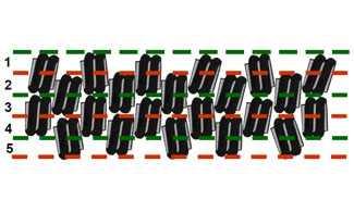
Thus, we have not detected any regular organization of nucleosomes, but our results indicate that chromatin in native chromosomes is organized forming well-defined plates. To justify the small thickness observed for plates, we have suggested that it is very likely that there is an interdigitation between the successive layers. We think that the resulting compactness is necessary for the safe transfer of the genomic DNA to the daughter cells during mitosis. The thin-plate model provides a completely new interpretation of the metaphase chromosome structure. Our model gives the physical basis to explain easily the formation of chromosome bands which are studied in the cytogenetic analyses.
The images of TEM, SEM, and of cryo-electron microscopy and electron tomography have been obtained in the Servei de Microscòpia of UAB, the AFM images and the force spectra in the Serveis Científico-Tècnics of UB, and the images of polarizing microscopy in the Servei de Tractament i Anàlisi d’Imatges of UAB.
Pablo Castro-Hartmann, Isaac Gállego, Maria Milla, Silvia Caño, Juan Manuel Caravaca, Joan-Ramon Daban
References
"Dense chromatin plates in metaphase chromosomes". I. Gállego, P. Castro-Hartmann, J.M. Caravaca, S. Caño i J.R. Daban. European Biophysics Journal, 38 (2009) 503-522.
"Irregular orientation of nucleosomes in the well-defined chromatin plates of metaphase chromosomes". P. Castro-Hartmann, M. Milla i J.R. Daban. Biochemistry, 49 (2010) 4043-4050.