Fear-processing mechanisms of the brain documented
24/07/2015
The researchers systematically analysed the studies conducted up to now that measure brain activity during the process of fear conditioning through functional magnetic resonance (fMRI). Their findings show that in this process there is a robust pattern of activation in a set of regions of the brain, which could make up the “fear network”.
These findings will give us a greater insight into the transition between normal and pathological fear and, in the long term, will enable us to optimise the physiopathological models for tackling anxiety disorders.
The usual experimental approach to studying fear-learning mechanisms makes use of (Pavlovian) fear conditioning. In this model, an initially neutral stimulus (e.g. seeing a dog) produces a fear response when paired with an aversive stimulus (e.g. being bitten by a dog). The incident with the dog transforms the normally neutral sight of any other dog into a stimulus that produces fear.
The fMRI technique allows us to see the brain regions that are carrying out a particular task, such as fear learning. In this case, the team of researchers analysed a total of 27 studies on fear conditioning made between 1998 and 2013, using fMRI, and involving a total of 677 healthy adult participants.
This type of work is of great importance to the evaluation of existing evidence on a topic, as it comprises a larger number of observations: meta-analysis is of greater statistical power than the studies included in it.
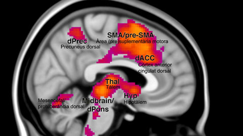
In this case, ground-breaking meta-analysis techniques were used, which enabled the study of brain deactivation as well as activation. "In the field of neuroimaging, functional deactivations of brain regions can be just as informative as functional activation for understanding the neural substrates of complex mental activities like processing emotions", explains Miquel Àngel Fullana, the researcher who coordinated the study, from the Department of Psychiatry and Legal Medicine of the UAB and Hospital del Mar.
The scientists found a common pattern in all the studies analysed: the coordinated activation of different brain regions distributed anatomically. “Foremost among the regions that participate in fear conditioning are the "cingulofrontal cortex" regions, including the insula and the dorsal anterior cingulate cortex. Moreover, this brain network has been linked to interoception, the sense of the physiological condition of our body, explains Miquel Àngel Fullana.
Learning to identify and respond to threat signals is critical to survival, as it generates a physiological and behavioural response that allows the threat to be avoided or faced. However, when this process becomes dysregulated and elicits fear responses to innocuous events, it can lead to anxiety disorders. “The way in which fear and anxiety are experienced and the link to this neural network have not been sufficiently explored in the neuro-scientific study of patients with anxiety disorders, and therefore represent a novel research avenue", concludes Dr Fullana.
Approximately 15% of the population will experience an anxiety disorder at some point in their life, though the group of persons suffering from severe anxiety disorders is smaller.
This research was carried out by a team of researchers from the Department of Psychiatry and Legal Medicine of the UAB, the Anxiety Unit, Institute of Neuropsychiatry and Addictions, MAR Healthcare Park, and the Hospital del Mar Institute for Medical Research (IMIM).
Reference article
Fullana MA, Harrison BJ, Soriano-Mas C, Vervliet B, Cardoner N, Àvila-Parcet A, Rauda J. Neural signatures of human fear conditioning: an updated and extended meta-analysis of fMRI studies. Mol Psychiatr 2015. Doi:10.1038mp.2015.88